Specific Integrin Labeling in Living Cells Using Functionalized Nanocrystals
20-Aug-2007
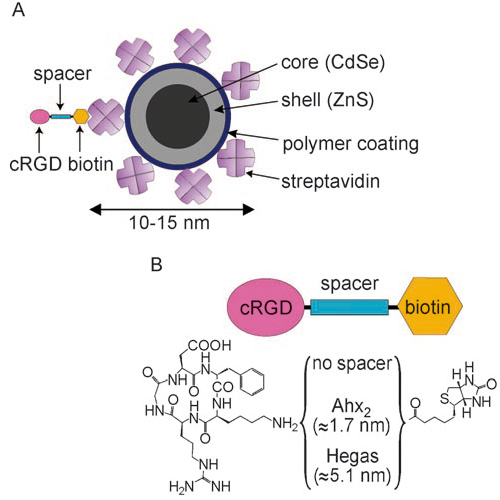
Quantum dot (QD) bioimaging has recently been described as the most exciting new technique to emerge from the collaboration of physicists and biologists. This new molecular imaging technology is based on the selective fluorescent labeling of biological molecules. QDs are fluorescent nanocrystals showing a wide-range absorption spectrum (400–650 nm) and a narrow, symmetric emission spectrum. By choosing the appropriate QD size, the emission wavelength can be tuned even to the near infrared. This allows the QD signal to be clearly distinguished from the cell autofluorescence background. Finally, the stability of QDs against oxidation and photobleaching makes them highly suitable for single-molecule labeling compared to conventional dyes. Based on these unique spectral, physical, and chemical properties, QDs are extremely useful as a technique to “light up” biological events. However, besides all their advantages, QDs do not offer a continuous fluorescence signal as they exhibit a statistical blinking behavior. This feature hampers automated image processing and might adulterate the quantitative parameters that are extracted from the trajectories of such blinking particles.


