

Dying cells expose a nuclear antigen cross-reacting with anti-PD-1 monoclonal antibodies
11-Jun-2018
Scientific Reports, volume 8, Article number: 8810 (2018), https://doi.org/10.1038/s41598-018-27125-6
Scientific Reports, online article
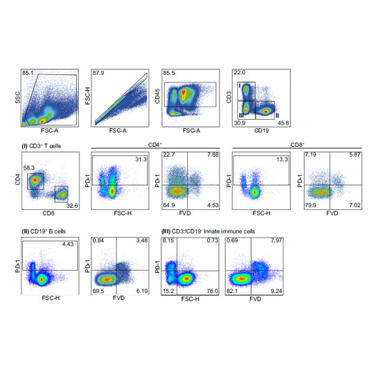
Checkpoint molecules such as programmed death 1 (PD-1) dampen excessive T cell activation to preserve immune homeostasis. PD-1-specific monoclonal antibodies have revolutionized cancer therapy, as they reverse tumour-induced T cell exhaustion and restore CTL activity. Based on this success, deciphering underlying mechanisms of PD-1-mediated immune functions has become an important field of immunological research. Initially described for T cells, there is emerging evidence of unconventional PD-1 expression by myeloid as well as tumor cells, yet, with cell-intrinsic functions in various animal tumor models. Here, we describe positive PD-1 antibody staining of various murine immune and tumour cells that is, unlike for T cells, not the PD-1 receptor and restricted to cells with low forward scatter characteristics. Based on flow cytometry and various approaches, including two established murine anti-PD-1 antibody clones, CRISPR/Cas9 genome editing and confocal imaging, we describe a staining pattern assigned to a nuclear antigen cross-reacting with anti-PD-1 monoclonal antibodies. Lack of PD-1 expression was further underlined by the analysis of PD-1 expression from B16-F10-derived 3D cultures and ex vivo tumours. Thus, our data provide multiple lines of evidence that PD-1 expression by non-T cells is unlikely to be the case and, taking recent data of PD-1 tumour cell-intrinsic functions into account, suggest that other antibody-mediated pathways might apply.