

Common Fibril Structures Imply Systemically Conserved Protein Misfolding Pathways In Vivo
23-May-2017
Angew. Chem. Int. Ed., Volume 56, Issue 26, Pages 7510-7514, https://doi.org/10.1002/anie.201701761
Angew. Chem. Int. Ed., online article
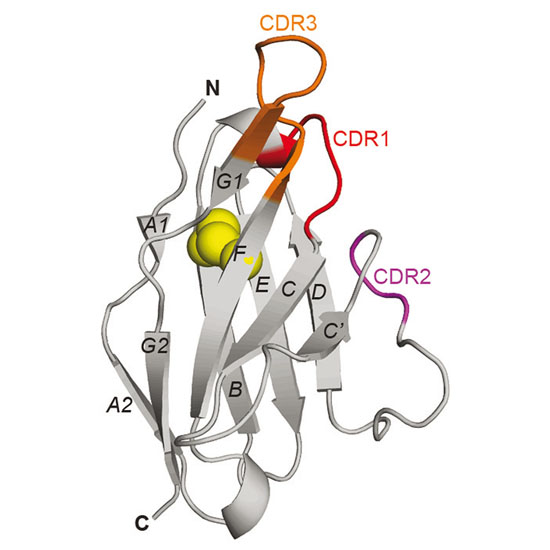
Systemic amyloidosis is caused by the misfolding of a circulating amyloid precursor protein and the deposition of amyloid fibrils in multiple organs. Chemical and biophysical analysis of amyloid fibrils from human AL and murine AA amyloidosis reveal the same fibril morphologies in different tissues or organs of one patient or diseased animal. The observed structural similarities concerned the fibril morphology, the fibril protein primary and secondary structures, the presence of post‐translational modifications and, in case of the AL fibrils, the partially folded characteristics of the polypeptide chain within the fibril. Our data imply for both analyzed forms of amyloidosis that the pathways of protein misfolding are systemically conserved; that is, they follow the same rules irrespective of where inside one body fibrils are formed or accumulated.